Exploring the Golgi Tendon Organ and Muscle Spindles’ role in the deadlift.
DISCLAIMER
Anatomy, physiology, AND pathophysiology lie ahead!
Want to skip it and head straight to the summary?
CLICK HERE
Want my final thoughts?
CLICK HERE
Introduction
We’ve all been there. Metallica blasting out over the airwaves, barefoot to reduce the length the bar has to travel, and that feeling of two sharp pencils being shoved up your nose as a result of snorting that fresh, unopened, jar of smelling salts that’s potent enough to revive the dead. What better way to prepare for a Deadlift PR?!
You imagine you’re Teddy Sherringham at the Nou Camp, Barcelona 99′. It’s the 89th minute and you’re about to save the day! You then remember the cue, ‘it’s not a pull, it’s a leg press through the floor’!
You break the tension on the bar, then rip that bad boy up like there’s no tomorrow! Nothing’s gonna stop you locking it out. Boken spine? You’ll take it. All day long if it means you smash the lift, upload to Insta, and impress all the tens of followers that are sat on the toilet with nothing else to watch.
All of a sudden… you did it. Your mind ascends to a godlike ethereal realm where you feel unstoppable. You can now pull anything, press everything. Hell, with Van Halen’s earsplitting guitar riffs pounding through your brain, you could run faster than a gazelle straight into the path of a 50 cal and it’d wouldn’t even leave a flesh wound! You ain’t got time to bleed.
You’re Freddie Mercury on steroids. Pisarenko himself would bestow upon you the title of the manliest of all men!
But among all the rushing endorphins and elation, you can’t quite figure why the heck your legs shook so much?!
Well, don’t fear! We’re going to lean over and peak into the rabbit hole with this one. Firstly, let’s go over the billy basics.
MSK 101
Three types of muscle exist in the human body:
Cardiac muscle is found in the heart, making up its thick middle layer. It is responsible for the hearts pumping action. This is classed as involuntary, as we do not consciously control this. Involuntary muscles sustain long or near-permanent contractions.
Smooth muscle is found in the walls of internal organs and blood vessels. This, like cardiac muscle, is classed as involuntary. It’s responsible for all the contractions that occur in organs, such as controlling the peristaltic movement in the oesophagus and intestines.
Skeletal muscle is fairly self explanatory. It is classed as voluntary muscle, as we can consciously control this. For the purpose of this article, this is the one we are interested in.
Skeletal muscle produces force that contracts. It is connected to bone via tendons.
Skeletal muscle produces movement, maintains body posture, controls body temperature, and stabilises joints.
The muscle belly is composed of fascicles, which themselves contain between 20-80 muscle fibres (sometimes referred to as myocytes), each of which then contain thousands myofibrils.
Myofibrils are the contractile elements of the muscle, containing repeating units called sarcomeres. These will be briefly explored later in the article.

Connective tissue hold all these bundles together. There are three types:
Epimysium – surrounds the entire muscle.
Perimysium – surrounds each fasicle (i.e. bundle of muscle fibres/myocytes).
Endomysium – surrounds each individual muscle fibre.
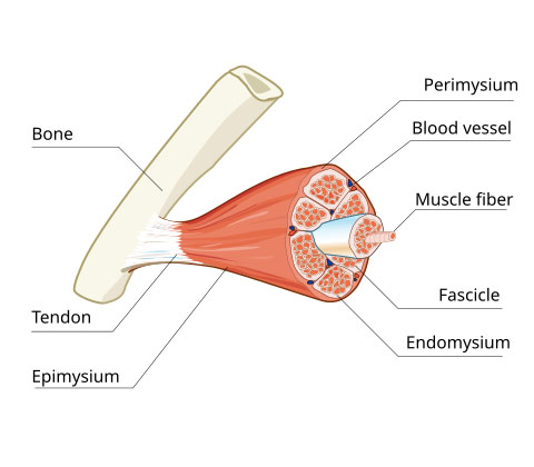
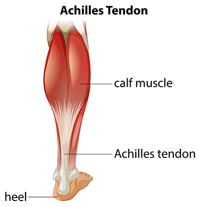
Tendons are on the end of our muscles. They act as the intermediary between muscle and bone. Muscle connects to tendons via the myotendinous junction (MTJ), which itself forms part of the fairly complex myotendinous unit.
The MTJ is composed of tendon fibres and terminal muscle fibres which create finger like projections that increase the surface area between tendon and muscle. This increased surface area acts to disperse the energy of the contracting muscle, thereby decreasing focal stress. In laymans terms, it functions to ‘spread out the load’.
Proprioceptors
To understand why we shake during a deadlift, we first need to understand the structure and function of two neurological sensory receptors (aka. proprioceptors), which are located in our muscle and tendon fibres.
1. Muscle Spindles
2. Golgi Tendon Organs (GTO’s)
Muscle Spindles sense STRETCH and SPEED (VELOCITY). They are located in the muscle belly, running parallel to the muscle fibres. Their main role is in the action of the stretch reflex, which will be covered soon.
GTO’s sense muscle TENSION (FORCE). They are located within the myotendinous junction (MTJ).
*Note – you may see the term ‘enthesis’ used to describe connecting interfaces. Whilst serving a similar function to the MTJ, an enthesis is actually the bit that connects tendons and/or ligaments to bone, otherwise known as the tendon-to-bone interface (TBI for short). Both the MTJ and TBI are illustrated below.

Muscle Spindles
Extrafusal muscle fibres form the outer bulk of the muscle, contracting and generating skeletal movement. They have an origin and insertion point on our bones, and have a fusiform (spindle) shape to them, hence the name extrafusal.
Intrafusal muscles fibres run parallel to the extrafusal muscle fibres. The sheath containing the fibres are also fusiform, hence the name intrafusal. They don’t share the same origin and insertion point that the extrafusal fibres. They are instead attached to the intramuscular connective tissue. Intrafusal muscle fibres, combined with their nerve supply, form the Muscle Spindle. See below.

Each end of the muscle spindle have contractile elements (actin and myosin) that form the crossbridges responsible for muscle contraction. This is the same mechanism responsible for contraction in extrafusal muscle fibres (aka. the sliding filament theory).
The centre of the muscle spindles however contain little to no contractile elements, instead it contains the nuclei of the muscle fibre which serve as the sensory unit of the spindle. There are two types of intrafusal fibres based on whether the nuclei are concentrated in a wide central portion (Nuclear Bag Fibres), or if they are aligned in a chain (Nuclear Chain Fibres).
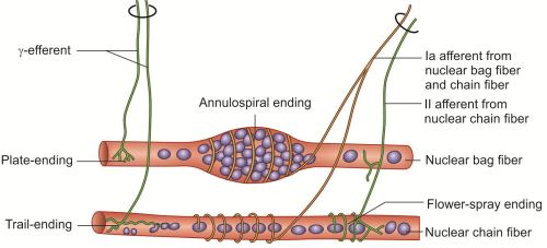
There are then two types of sensory (or afferent) nerve fibres that wrap around the spindle and are stimulated by the stretching of the mid-portion (i.e. the centre) of the muscle fibre:
Primary (Type Ia) fibres wrap around both Nuclear Bag and Nuclear Chain Fibres, and are considered to transmit information regarding velocity AND muscle length.
Secondary (Type II) fibres innervate one or both sides of the Nuclear Chain Fibres, and these are considered to only sense muscle length.
The stretching effect can occur either as a result of the lengthening of the extrafusal muscle fibres, OR contraction of only the muscle spindle end portions (i.e. the portions with actin and myosin).
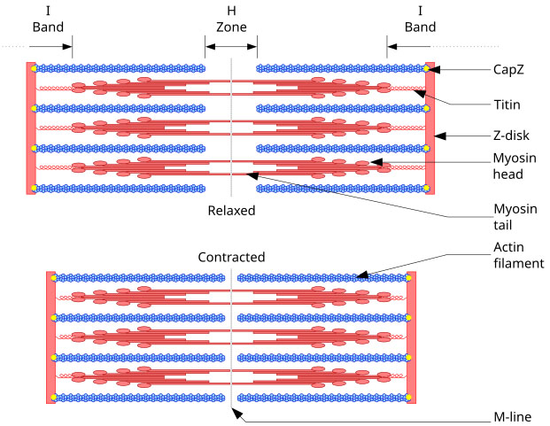
Now we may go into how contraction of muscle fibres occur (aka. the sliding filament theory) in a further article. For now, just remember that a single muscle fibre contains lots of tube like structures called myofibrils (see below).
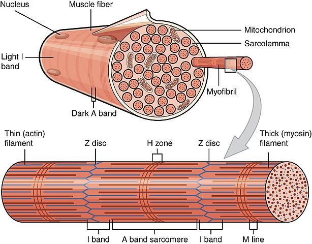
These myofibrils contain actin and myosin protein filaments, which form crossbridges with each other and ‘slide’ back and forth along the length of the myofibril – resulting in muscle contraction.
Titin (another protein filament) is also present in a myofbril, however this is considered to be an elastic protein rather than a contractile one. It functions as a molecular spring that is responsible for the passive elasticity of muscle. This can be seen in the image below.
As can be seen in the image by Pramanik, intrafusal muscle fibre ends are innervated by efferent nerve fibres (Gamma Motor Neurons to be precise) because they are contractile.
Extrafusal muscle fibres are also innervated by efferent nerve fibres, but these are Alpha Motor Neurons.
This distinction is worthy of note because it means that the contractile elements of the intrafusal muscle fibres receive seperate motor innervation. Which is perhaps a topic for another day!
The Stretch Reflex
The primary purpose of the stretch reflex is to oppose sudden changes in the length of the muscle. It does this to protect the muscle from potential damage as a result of sudden overstretching.
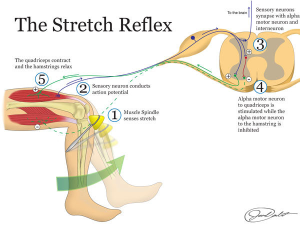
When the centre of the muscle spindle senses a sudden stretch, the afferent nerves (mainly the Primary Type Ia fibres) send this information straight to the dorsal column of the spinal cord.
This provides the shortest path of travel for the action potential to then synapse with an alpha motor neuron of the same muscle (remember, alpha motor neurons innervate the larger extrafusal muscle fibres).
Also remember that the centre of the muscle spindle (containing all of the sensory neurons) was initially excited by the change in length of the extrafusal muscle fibre.
The firing rate of the sensory nerves in the muscle spindle is dependent on the length, and speed of change in length, of the muscle fibre. Simply put, they sense if a stretch is TOO FAST and/or TOO FAR.
If the stretch is determined to be too fast or too far, then the alpha motor neurons fire action potentials down into the extrafusal muscle fibres, telling them to contract to prevent injury.
The opposite is true when there is no stretch applied to the muscle spindles, i.e. the extrafusal muscle fibres relax.
The stretch reflex is completely autonomous from the brain, as there are no higher cortical functions involved in this process.
Reciprocal Inhibition
We can see from the image above that in addition to the muscle spindle sending messages through to the motor nerve of the same muscle telling it to contract, it also branches off to synapse with an inhibitory interneuron which releases inhibitory neurotransmitters that decrease the excitation of the motor neurons to the antagonistic (or opposite) muscle, relaxing it.
That is to say that in the example above, when the quadriceps contract in response to muscle spindle activation, the hamstring will relax allowing the reflexive movement to occur without resistance from the hamstring.
*Geeky Side Note 1*
Monosynaptic & Polysynaptic Reflexes
Reflex arcs can be classed as monosynaptic (i.e one synapse), or polysynaptic (i.e. two or more synapses).
The stretch reflex is classed as monosynaptic. That is an efferent (sensory) neuron synapsing with an afferent (motor) neuron.
However reciprocal inhibition involves polysynaptic reflexes due to the addition of the interneuron that inhibits (relaxes) the antagonistic muscle.
That is to say that in the simplest form, the patella reflex (seen by tapping the tendon of the quadriceps) elicits the mono synaptic stretch reflex response, contracting the quadricep in response to the stretch. However, the reciprocal inhibition that causes the hamstring to simultaneously relax is considered to be polysynaptic.
i.e – the inhibitory interneuron involved in reciprocal inhibition receives the same action potentials from the initial efferent (sensory Type Ia fibres) in the stretch reflex, then it transmits inhibitory signals to the antagonistic muscle (the hamstring) via alpha motor neurons, so that it relaxes allowing the quadriceps to contract without resistance of hamstrings.
N.B. Interneurons can distribute signals segmentally throughout the spinal cord.
They be either inhibitory, OR excitatory.
*Geeky Side Note 2*
Consciously Overriding the Stretch Reflex
Due to the presence of interneurons, the central nervous system (the brain) can consciously inhibit the stretch reflex.
This is because voluntary upper motor neurons can travel to the spinal level involved in the stretch reflex response and stimulate or inhibit the lower motor neuron via the interneurons.
So in the patella reflex, we can tell our leg to kick out or stay put. This however, would then not be a true reflex as it is not involuntary.
*Geeky Side Note 3*
Using the Stretch Reflex to Identify an Upper Motor Neuron Lesion
As physios we can leverage reciprocal inhibition and use it to our advantage when it comes to identifying the presence of an upper motor neuron lesion.
We can use the Babinski reflex as an example. Here, we scrape across the sole of the foot, starting from the outside of the heel through to the base of the first metatarsal. In a developed pyramidal tract we expect to see the toes flex downwards, because this demonstrates inhibition of the toe extensors.
If we don’t see reciprocal inhibition of the toe extensors (that is to say, we see the great toe curl up, with or without the others fanning out), then we record a positive Babinski sign and we can deduce that either:
- An upper motor lesion exists somewhere along the pyramidal tract that is compromising the inhibitory action potentials (nerve signals) at vertebral level of the reflex.
- The pyramidal system is underdeveloped, as is seen in that of babies.
Golgi Tendon Organs (GTO’s)
You may remember from the moment arms in powerlifting article that force = mass x acceleration. A force can cause an object to accelerate, decelerate, deform, or rotate. Force is a vector quantity, meaning it has both magnitude (i.e. distance) and a direction of travel.
Common types of force include: Frictional Force, Gravitational Force, Spring Force, Normal Force, Magnetic Force, Electrical Force, Air Resistance Force, Applied Force and Tension Force.
Tension is defined as the force transmitted transmitted axially along an object when pulled by forces acting from opposite sides. It is the opposite of compression.
In our case, we can see that the forces acting from opposing sides are the contractile forces from one end of the muscle to the other – thus creating tension in the tendon.
GTO Function
GTO’s sense this tension force within a tendon. They are responsible for autogenic inhibition (also referred to as the inverse myotatic reflex or the golgi tendon reflex). This is a protective reflex, utilising a negative feedback loop, that prevents muscles from exerting more force than their tendons and bones can handle.
Structurally GTO’s are encapsulated bundles of collagen that are innervated by Type Ib afferent motor fibres, which are thinner and transmit signals faster than the Type Ia fibres found in the muscle spindle. These fibres are activated when the tendon is subjected to high levels of force, where they subsequently inhibit muscle activity to prevent injury.
The function of the GTO can therefore be considered the opposite of the muscle spindle, which serves to produce muscle contraction.
So, try to stick with me now! Here we go…
GTO’s detect the degree of tension in the tendon that is generated by muscle contraction, with the purpose of preventing tendon avulsion.
Whenever GTO’s are stimulated by tension, they activate type Ib sensory fibres which send that information (via action potentials) into the Central Nervous System (CNS).
Type Ib fibres travel to the CNS via the dorsal root ganglion (DRG), then move into the dorsal horn of the spinal cord – where they then have two options because it then bifurcates (or splits).
When it does this, it synapses on two different interneurons. See below:

One interneuron is inhibitory, which connects with an alpha motor neuron that travels to the extrafusal muscle fibres of the agonist.
The other interneuron is an excitatory (or stimulatory) interneuron, that connects with an alpha motor neuron that travels to the extrafusal muscle fibres of the antagonist (or opposite) muscle.
*Note – Motor Neuron’s reside in the anterior (or ventral) horn of the spinal cord, whereas Sensory Neurons reside in the posterior (or dorsal) horn of the spinal cord. You can see an example of this in the image above.
We can put all of this into context by using the bicep curl as an example. Imagine curling 200kg (i.e. a very heavy weight).
If the bicep is contracting so much that it’s at risk of pulling the tendon off the bone (an avulsion fracture) – do you want the bicep to keep contracting? No, you definitely don’t!
You want the muscle to do the opposite of contract – you want it to relax. That is to say, you want inhibition of the muscle.
So, in the autogenic inhibition reflex (i.e. the golgi tendon reflex/inverse myotatic reflex) – the nerve signals, generated by tension on the Golgi tendon organ residing in the bicep tendon, travel across the type Ib sensory fibres to the CNS via the DRG, into the dorsal horn of the spinal cord, THEN the signals pass through to the inhibitory interneuron.
This synapses with an alpha motor neuron that travels to the muscle (in our case, the bicep), and sends messages to the bicep telling it to relax – thus taking tension off the bicep tendon, thereby decreasing the risk of an avulsion fracture. See below:
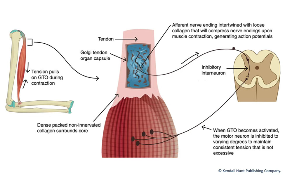
Now whenever we shorten and relax a muscle, we need the antagonist muscle to contract to pull the joint in the opposite direction to aid in taking tension off the tendon.
In the example above, the antagonist is the tricep as this extends the elbow. You can see the bifurcation of the Type Ib fibres in this image, however they are referring to the hamstring as the antagonist as the example on display is knee extension (not bicep flexion).
So, autogenic inhibition can lead to inhibition of the contracting muscle AND stimulation of the antagonistic muscle.
Interneuron Function
You might then ask, HOW do the interneurons act as inhibitory or excitatory? Well…
We’ll do a deepish dive into nerve conduction in a future article. But for now, the answer is that it all depends on the type of neurotransmitters that are released in the synapse.
The neurotransmitter that’s released from the inhibitory neuron tends to be glycine. Glycine generates inhibitory post synaptic potentials, or I.P.S.P’s for short. This means they cause negative ions to rush in, or positive ions to rush out of the cell – making the inside of the cell very negative (thus, hyper polarising it).
The neurotransmitter that’s released from the stimulatory interneuron tends to be glutamate. Glutamate generates excitatory post synaptic potentials, or E.P.S.P’s for short. This means they cause cations (positively charged ions) to flow into the cell, depolarising it which leads to action potentials.
So, Why Do My Legs Shake When I Pull A Heavy Deadlift?
Whoa! That was a deep-ish dive into the stretch reflex and the golgi tendon reflex.
But hopefully we now understand the two statements below. We can then apply these to the deadlift:
- The stretch reflex, combined with reciprocal inhibition serves to protect a muscle from potential damage by sensing the length of stretch on the muscle, and the speed at which that stretch occurs. If the muscle spindles determine the stretch to be too long or too fast, they send excitatory signals to the agonist muscle telling it to contract – thus protecting the muscle from overstretching. Simultaneously, signals pass through to the antagonist muscle telling it to relax so that the agonist doesn’t have to produce more force to overcome any muscular contraction of the antagonist.1
- The Golgi tendon organ senses tension in the tendon of the agonist muscle. If this is determined to be too high (risking tendon avulsion), inhibitory signals are sent through to the agonist telling the extrafusal muscle fibres to relax. Simultaneously, motor signals are sent through to the antagonist telling it to contract, in an attempt to further reduce tension on the tendon of the agonist.
The DEADLIFT
The deadlift is generally considered to have three phases:
Lift off – this is when the force is applied to the bar to pull it off the floor.
Mid pull – this is when the bar is located immediately above the knees
Lock out – this is when the trunk reaches the vertical position with the bar positioned at its highest point.
LIFT-OFF
Hamstrings
The hamstrings, particularly biceps femoris (BF), have been shown to exhibit less activation in the lift-off position compared to mid pull and lock out.
It is proposed that this is because the BF is in an unfavourable position due to the length-tension relationship, especially in relation to the hip joint when the knee and hip joint are more flexed and the BF more elongated.1
Quadriceps
Load on the quadriceps, particularly rectus femoris (RF) has been shown to be at its greatest during the lift off phase, and at its lowest during the mid pull phase.
This is said to occur because the smaller angle of the knee and hip during lift off would request higher demand of the RF, while the hamstrings act as knee stabilisers. The RF muscle is also a hip flexor and would only need to act on the knee joint in this position too.1
MID-PULL
Hamstrings + Quadriceps
Load on the hamstrings, particularly the BF, is at its highest during mid pull. The increased activation is said to be the result of a position in which BF is acting as a hip extensor at an optimal angle.
Additionally, the amplitude of the knee in this position is closer to maximum extension than the hip joint, generating a higher possibility of tension in BF as the hip reaches maximum extension. The maximum torque in isometric hip extensions is higher when BF is in a position more elongated by knee extension, which corresponds to the mid-pull position.1
SHAKING IN THE DEADLIFT THEORY
Yes – that is all the following is. A theory.
This is because I can find no empirical data that provides clear reasoning as to what mechanism is at play to cause the shaking in the deadlift. If any readers can find such evidence – PLEASE get in touch.
I am simply pulling together data from academia, known principles such as reciprocal and autogenic inhibition, and my own anecdotal evidence of over 20 years deadlifting to draw a reasonably informed conclusion as to what is likely happening. Hence, I MIGHT BE WRONG!
Clonus
Now before I put forward my theory, I will say that I’ve heard many a bro-scientist refer to the shaking/jerkiness as clonus. I humbly disagree. This is why…
Clonus is an abnormal reflex response that involves involuntary and rhythmic muscle contractions. It is often accompanied by hyperreflexia and is indicative of an upper motor neuron lesion, much like the Babinski sign.
Although clonus may visually appear identical to the shakiness/jerkiness that can occur in a max effort deadlift, the underlying cause of such actions are not.
In literature, there are currently two theories that try to explain the underlying mechanisms behind clonus:
- It’s a self-perpetuating reactivation of peripheral muscle stretch circuits, with each beat producing the next.
- There’s an initial appropriate external stimulus that leads to activation of the stretch reflex circuit, followed by a central signal which commands the muscles to continue to produce that motor response in the absence of an appropriate stimulation of the stretch reflex.2
Hyperexcitability in muscle stretch circuits is produced when there is less tonic inhibition of motor neurons involved in the monosynaptic stretch reflex. Think back to those good old monosynaptic and polysynaptic reflexes. For further reading on the difference between tonic and phasic inhibition, I would suggest exploring this article.
In short, tonic firing refers to a sustained response, which activates during the course of the stimulus. Phasic firing refers to a transient response with one or few action potentials at the onset of stimulus followed by accommodation.3
Hyperexcitability can occur when there is a lesion to descending motor nerves, predominantly the dorsal reticulospinal pathway, which can occur anywhere from the cortex to the spinal cord.
The inhibitory dampening effect of these descending nerves on alpha and gamma motor neurons is removed, leading to a hyper excitatory state in the muscle stretch reflex circuit.
Clonus is therefore considered to be a manifestation of an upper motor neuron pathology, which explains why other signs of hyperreflexia generally accompany it.2
SHAKING IN THE DEADLIFT
I’ve hopefully convinced you now that if you shake during a deadlift, it doesn’t mean you have an upper motor neuron lesion.
Proposed mechanism of the deadlift tremor:
I think this can most definitely (dude), be attributed to either the stretch reflex or Golgi tendon reflex – or potentially both. Below, I propose this to mostly be the result of a battle of the Golgi tendon organs.
Theory:
We know that shaking occurs mostly during mid pull. We have also ascertained that tension on the hamstring tendons are at their highest during the mid pull phase, and tension on the quadriceps are at their lowest during mid phase.1
At max effort (RPE 11!), this tension could cause the Golgi tendon reflex to inhibit muscle activity to the hamstring, thus preventing an avulsion fracture, whilst simultaneously firing motor signals to the antagonist (the quadriceps) telling it to activate and shorten, thus reducing further tension on the hamstring tendons.
This results in increased tension on the quadriceps/patella tendon. This would similarly then result in the Golgi tendon reflex in the quad/patella tendon sensing all that tension and inhibiting quadricep activation in an attempt to prevent an avulsion fracture. Simultaneously, the reflex arc would stimulate muscular contraction of the hamstrings to further reduce tension off the quadricep/patella tendon.
What follows is a battle of the Golgi tendon reflex in both the hamstrings and quadriceps, which would likely present as shaky/jerkiness in the legs, reducing at lock out once tension on the tendons has somewhat decreased.
DISCUSSION
I’m going do that thing that every student at university does when writing a research article… I propose that further research needs to be undertaken if we are to fully understand the physiology behind why we shake during a deadlift.
Most studies out there don’t appear to test the gluteus maximus (GM) during a conventional deadlift. I think the addition of monitoring muscular activity in the GM, AND the semi-tendinosis (ST) and semi-membranosus (SM), during each portion of the deadlift could help ascertain whether the addition of the GM in terminal hip extension has a role to play with the cessation of the shaking, and/or reduction of tension on hamstring tendons.
If it was proved that the GM takes over the primary role of hip extension in this phase, this could provide further reasoning as to why the shaking stops at lock out. This theory would however be compromised if hamstring and quadriceps activity were, as per previous studies1, to show no change (i.e. no drop) in electromyographic (EMG) activity towards lock out.
EMG
Furthermore, I recognise that surface EMG testing has limitations in this context. Mainly because when a joint angle changes (thus changing the muscle fibre length), the diameter of the muscle fibre changes because muscle volume is practically a constant (n.b. – volumetric strain-energy potential has been shown to reduce muscle volume by 2–4% during contraction of fully active parallel muscle fibres6). Though for all intents and purpose, the volume remains relatively constant throughout. Due to the change in changing geometry of the muscle being monitored, it makes it difficult to accurately monitor the recruitment pattern among changing joint angles.
To aid in visualising this, imagine you were happily minding your own business monitoring the flow of oxygen atoms in a slippery water snake toy, then someone comes along and squeezes the slippery snake, pushing all of the water (hence, oxygen atoms) up and off the sensor! This would compromise the accuracy of your results.
Okay, maybe there’s a better example out there. But hopefully you get the point 😊
Intramuscular EMG may therefore provide more accurate results, however this would likely be too invasive for the participant and highly challenging for the assessor. Nonetheless, it remains an option.
MONITOR MORE MUSCLES
Furthermore, in the majority of studies referenced, only one single hamstring muscle has been monitored. This has its limitations in terms of being able to draw accurate conclusions on what occurs in all three hamstrings during knee flexion/extension and hip flexion/extension.
To elaborate, the semi-tendinosis (ST) and semi-membranosus (SM) have more distal attachments compared to the long and short the of the BF. This suggests that the ST and SM have more advantageous moment arms for production of force in knee flexion, compared to that of the BF.7
Due to the above, there may actually be functional differences among individual hamstring muscles in hip extension and knee flexion. This means that monitoring just one hamstring has it’s limitations in terms of accuracy.
MUSCLE GEOMETRY
Lastly, in order to fully understand the tension forces acting upon each muscle during the deadlift, there should be recognition of muscle architecture of each individual muscle being tested and the effect that has on the force-velocity relationship. That is to say that fusiform muscle has been found to be more effective in muscle contraction velocity, whereas pennate muscle has been found to be better in muscle force generation.

This fact is likely due to fusiform muscles allowing a greater range of motion – and we know that fibre length is an important determinant of the quantity of contraction possible in a muscle.
Pennate muscle fibres however are diagonally orientated, which maximizes the muscle’s force potential. Additionally, more muscle fibres fit into the muscle compared with a similarly sized fusiform muscle.5
We know that all four hamstrings are fusiform, with varying degrees of fibre length and physiological cross sectional areas. The quadriceps are however considered to be bipennate.8 Could this geometry have a role in the distribution of tension forces during each phase of the deadlift? Probably! But all this thinking is starting to make my head hurt, so I’m ending it here 🙂
CONCLUSION
As you can see, and as with most studies – method of testing, accuracy of testing (i.e. inter and intra-rater reliability), and subtle differences in participant anthropometry can all affect the results. Ideally, we could throw billions at this and produce the most accurate results ever, meaning that we could draw definitive conclusions as to what exactly occurs when we shake during a deadlift.
… But ain’t nobody gonna do dat!
I therefore propose that muscular tension in each tendon of SM, ST, BF, RF and GM, gastrocnemius and erector spinae should be monitored throughout each portion of the lift using electrical impedance, as this has been found to be a good measure of tendon tension.4
FINAL THOUGHTS
Urgh, I’m not an academic and now my head REALLY hurts. I think someone far more intelligent than me can now take the reins on this one 🙂
At the end of the day, does anyone really care?! Let’s just embrace the shake, and lift some heavy ass weight!
I’m off to get a beer, or two 🍻
If you have any comments, compliments or complaints, please write them below.
Oh, and if you need a bar to deadlift with… you can help the site by purchasing one here.
References
IMAGES
Lim, HoTae & Choi, In & Hyun, Sang hwan & Kim, Hyesoo & Lee, Gabsang. (2021). Approaches to characterize the transcriptional trajectory of human myogenesis. Cellular and Molecular Life Sciences. 78. 1-14. 10.1007/s00018-021-03782-1.
Hariadhi. (2024) Skeletal Muscle. Available at: https://commons.wikimedia.org/wiki/File:Skeletal_muscle_svg_hariadhi.svg
BRGFX. (2014) Achilles Tendon. Available at: https://www.freepik.com/free-vector/medical-infographic-achilles-tendon_11253877.htm
Bianchi, E., Ruggeri, M., Rossi, S., Vigani, B., Miele, D., Bonferoni, M. C., Sandri, G., & Ferrari, F. (2021). Innovative Strategies in Tendon Tissue Engineering. Pharmaceutics, 13(1), 89. https://doi.org/10.3390/pharmaceutics13010089
Pramanik, D. (2015). Principles of Physiology. 5th edn. (no place):Jaypee Brothers Medical Publishers
OpenStax. (2015) Organization of Muscle Fiber. Available at: https://commons.wikimedia.org/wiki/File:1002_Organization_of_Muscle_Fiber.jpg
Richfield, D. (2007). Sarcomere. Available at: https://commons.wikimedia.org/w/index.php?curid=2264027
Winter, J.C. (2015). The Stretch Reflex. Available at: https://content.byui.edu/file/a236934c-3c60-4fe9-90aa-d343b3e3a640/1/module9/readings/somatic_reflexes.html
Winter, J.C. (2015). The Golgi Tendon Reflex. Available at: https://content.byui.edu/file/a236934c-3c60-4fe9-90aa-d343b3e3a640/1/module9/readings/somatic_reflexes.html
Wilson, P. (2014). Anatomy of Muscle. (no place):Elsevier
ARTICLES
- Moreira, V.M., Lima, L.C.R.d., Mortatti, A.L., Souza, T.M.F.d., Lima, F.V., Oliveira, S.F.M., Cabido, C.E.T., Aidar, F.J., Costa, M.d.C., Pires, T., et al. (2023) Analysis of Muscle Strength and Electromyographic Activity during Different Deadlift Positions. Muscles, 2(2):218-227. Available at: https://doi.org/10.3390/muscles2020016
- Zimmerman, B., Hubbard, J.B. Clonus. ( 2023) Treasure Island (FL): StatPearls Publishing. Available at: https://www.ncbi.nlm.nih.gov/books/NBK534862/
- Wang, L., Liang, P. J., Zhang, P. M., & Qiu, Y. H. (2014). Ionic mechanisms underlying tonic and phasic firing behaviors in retinal ganglion cells: a model study. Channels (Austin, Tex.), 8(4), 298–307. Available at: https://www.ncbi.nlm.nih.gov/pmc/articles/PMC4203731/
- Suganuma, J., & Nakamura, S. (2004). Measurement of tension of tendon tissue based on electrical impedance. Journal of orthopaedic science : official journal of the Japanese Orthopaedic Association, 9(3), 302–309. Avaiable at: https://doi.org/10.1007/s00776-004-0782-7
- Yanagisawa, O., & Fukutani, A. (2020). Muscle Recruitment Pattern of the Hamstring Muscles in Hip Extension and Knee Flexion Exercises. Journal of Human Kinetics, 72, 51–59. Available at: https://doi.org/10.2478/hukin-2019-0124
- Ryan, D.S., Domínguez, S., Ross, S.A., Nigam, N., Wakeling, J.M. (2020). The Energy of Muscle Contraction. II. Transverse Compression and Work. Frontiers in Physiology. Available at: https://www.frontiersin.org/journals/physiology/articles/10.3389/fphys.2020.538522/full
- Herzog, W., Read, L.J. (1993). Lines of action and moment arms of the major force-carrying structures crossing the human knee joint. Journal of Anatomy. Available at: https://www.ncbi.nlm.nih.gov/pmc/articles/PMC1259832/
- Takeda, K., Kato, K., Ichimura, K. & Sakai, T. (2023) Unique morphological architecture of the hamstring muscles and its functional relevance revealed by analysis of isolated muscle specimens and quantification of structural parameters. Journal of Anatomy, 243, 284–296. Available at: https://doi.org/10.1111/joa.13860

Whilst not writing for FGUK, Tim works as a Physiotherapist, Personal Trainer and is a Retired Ammunition Technician with the British Army. In his spare time Tim enjoys engaging in a whole variety of sports, spending considerable time with his little rascal of a dog, relaxing with his friends and family, but most of all.. geeking out on all things fitness!


